Day :
- Colorectal surgery case reports,Cardiology case reports,Gastroentrology case reports,Neurology case reports,Pulmonology case reports,Anaesthesiology case reports,Surgery case reports,Case reports on adverse drug reactions
Session Introduction
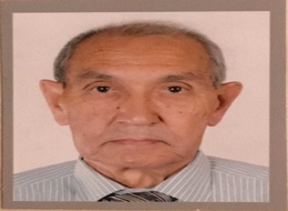
Karimbayev Kidirali
International Kazakh-Turkish University, Kazakhstan
Title: Late and rare complications of urolithiasis: Paranephric abscess and renal replacement lipomatosis

Biography:
Karimbayev Kidirali graduated from Tashkent Medical Institute as a Bachelor of Medicine in 1969, worked in regional hospital in Uzbekistan as an Anesthesiologist during 1969-1976 and in the year 1977 specialized as an Urologist. He earned Post-graduate degree in Urology at the Central Post Graduation Institute (Moscow 1977-1980) and worked as an Assistant Professor at the Department of Urology of Tashkent Medical Institute (1980-1993), then Head of Department of Urology of Republic Hospital (Tashkent, Uzbekistan). Since September 2013, he is working as Professor of Urology in Medical School of International Kazakh-Turkish University. In 1981, he completed his Dissertation Research Work “Transurethral resection in diagnostics of prostatic cancer” and was awarded with the degree of PhD and in 2002 - Dissertation Research Work “Modern methods in the treatment of long and complicated urethral strictures” Moscow with degree of Doctor of Medicine. He has more than 60 articles which were published in republican and international journals. Also, he is a Translator of the “Robbins Basic Pathology”. He translated the book from English to Kazakh language to give Kazakh Medical Students opportunity to study foreign Medical literature.
Abstract:
Background: We present a case of 62 year-old Kazakh women who for a long period of time suffered from back pain, weakness (has been treated with osteochondrosis), and was not aware about her kidney stones. She had never had hematuria or renal colic and urgently was admitted in a severe condition with large palpable abdominal mass.
Methods: Physical examination, laboratory data, radiology of the urinary tract, surgical treatment, histology, medical follow-up.
Results: On physical examination: The lumbar region on the right side is oedematic, hyperemic, and sharply painful on palpation. Oedema spreads down into iliac region and suprapubic area; and medially – to the median line with involvement of the umbilical ring. MRI of the urinary tract: The size of the right kidney is reduced; shadow of multiple stones seen in; the parenchyma is thinned. Beginning from the lower pole of the right kidney up to the pelvic cavity – shadow of a liquid mass with heterogenous density, measuring 21 x 8.4 cm is determined.
Surgical Procedure: Lumbotomy, lower paranephral abscess drainage. Later – right nephrectomy.
Histology: Renal replacement lipomatosis, severe renal tubular dystrophy.
Conclusions: Nontreated urolithiasis may lead to loss of the kidney function and development of life-threatening complications like paranephric abscess and renal replacement lipomatosis.

Biography:
Tony Ze Li is a General Surgical Registrar at The Canberra Hospital with a keen interest in Vascular Surgery and Cardiothoracic Surgery. He was a graduate from the prestigious Australian National University and has stayed locally to work in the Australian capital region since then. He has also done cardiac surgical placements in Texas Heart Institute in Houston, Texas. He is particularly interested in the endovascular aspects of vascular surgery based on the combination of both the surgical finesse of the procedures as well as the ever changing technology involved in this field. Before his career in Medicine, he has attained university degrees in both Biomedical Science and Biotechnology Commercialization.
Abstract:
Background: Endovascular aneurysm repair (EVAR) has been a long-stay surgical option to treat abdominal aortic aneurysms (AAA), both contained and ruptured. Endoleaks, defined as persistent flow in the aneurysm sac extrinsic to the endograft, is the most common complication. Type II endoleak (T2EL) results from collateral aortic branches (lumbar arteries, inferior mesenteric artery) flowing retrograde into the sac. It is often considered the Achilles' heel to EVAR due to the controversies in its timing of diagnosis and management. It’s accepted that T2EL are slow growing, could expand the aneurysm sac, and carry a small but significant risk of aneurysm rupture.
Case Presentation: This case reports an early post-EVAR complication due to T2EL. Sixty six (66) years old gentleman was brought to casualty hypotensive, minimally responsive with a rigid and tender abdomen. CT angiography revealed a ruptured AAA contained within the retroperitoneum. The patient subsequently underwent an uncomplicated EVAR procedure. However one hour post procedure in ICU, he was found to be suddenly in haemorrhagic shock. Exploratory laparotomy revealed 8 L of blood within the peritoneum. Aneurysm sac was opened and four strong bleeding lumbar arteries were found and oversewn. It’s hypothesized that vigorous T2EL from lumbar back- bleed, through the ruptured aneurysm sac led to ongoing bleeding into the retro-peritoneum and subsequently into the peritoneal space.
Conclusion: This case showed that T2EL are not all slow growing and innocuous. One should consider T2EL as a cause of a patient who’s acutely deteriorating post-EVAR. Early CT-angiographic imaging post procedure may be indicated in certain groups.
Shweta Dixit
Sir Gangaram Hospital, India
Title: The Etiology outcome and prognostic indicators in children with acute hepatic failure

Biography:
Shweta Dixit is presently pursuing a fellowship in Pediatric Gastroenterology from Sir Gangaram Hospital, New Delhi. She has done Diploma in Child Health and DNB in Pediatrics.
Abstract:
Objective: To study the etiology, outcome and prognostic indicators in pediatric acute liver failure (ALF).
Material & Method: A retrospective analysis of patients with ALF between 2010 and 2018 was performed. There were total 90 patients. Patients who survived with supportive therapy were designated as Group 1, while those who died or required liver transplantation were designated as Group 2.
Results: There were 90 children- 64 male, 26 females, median age eight years; range, one month-17 years were identified with fulminant hepatic failure. In Group 1 there were 53 (58.9%) patients and 37 (41.1%) patients in Group 2. The overall survival rate was 74%. Eleven (11) patients (12.2%) underwent liver transplantation, all are alive and well. The etiologies were: 60 (67%) infectious, 5 (5.5%) drug-induced, 5 (5.5%) Wilson disease and 3 (3.3%) autoimmune hepatitis. Overall acute viral hepatitis A is the most common cause. There were 16 (17.8%) cases which were of indeterminate etiology. Group 2 children had higher plasma bilirubin (median, 18.4 mg/dl versus 8.8 mg/dl), higher prothrombin time (median, 50.3 seconds versus 31.7 seconds) higher international normalized ratio (INR) (median, 4.9 vs 2.9) lower alanine aminotransferase (median, 1128 IU/L versus 2211 IU/L) and lower aspartate aminotransferase (median, 640 IU/L versus 1839 IU/L) levels on admission (P<0.005). Using receiver operating characteristic curves the cutoff values as predictor of the eventual failure of conservative therapy were serum bilirubin >13.6 mg/dl, prothrombin time >35.8 sec, INR >3.7, alanine aminotransferase ≤1913 IU/L, aspartate aminotransferase ≤ 1744 IU/L on admission.
Conclusions: Pediatric acute liver failure patients with high bilirubin, severe coagulopathy, lower alanine aminotransferase and lower aspartate aminotransferase level on admission are more likely to have poor prognosis. Early consideration of liver transplantation is of paramount importance in these patients.
Samih Abed Odhaib
Faiha Specialized Diabetes, Endocrine, and Metabolism Center,Iraq
Title: Preoperative diagnosis of a rare small bowel obstruction : Right paraduodenal herina: Case Report

Biography:
Samih Abed Odhaib has completed his Medical Education from Nahrain Medical School in 2001. He is a Fellow of Iraqi Board of Medical Specialization (Internal Medicine) FIBMS, Member of American College of Physician (ACP), Member of American Association of Clinical Endocrinologist (AACE). He is a Post-doctorate in Adult Endocrinology and is associated with Faiha Specialized Diabetes, Endocrine, and Metabolism Center (FDEMC) since September 2018. He was the Head of the Study Group that manage the case of Right Paraduodenal Hernia and the Manuscript Author.
Abstract:
The right paraduodenal hernia is an internal hernia of a rare type extends through the fossa of Waldeyer, caused by a congenital gut malrotation defect or trauma, infectious disease, iatrogenic trauma or mesenteric defect. The right is far less common than the left paraduodenal hernia (1:3), and three times more male predominant. A 67-year-old female with acute small bowel obstruction, with a diagnosis of right paraduodenal hernia done, preoperatively by imaging studies and confirmed during laparotomy with a good postoperative course and total resolution of obstructive symptoms. This will be the 168th case reported since Neubauer had published his first case in 1756.
- Case reports on diabetis , Cardiology case reports , Gastroentrology case reports ,Adverse drug reaction case reports ,Neurology case reports , Obstetrics and Gynecology case reports ,Health administration case reports ,Surgery case reports
Session Introduction
Hila Yariv
Reuth rehabilitation hospital, Israel
Title: An investigation into the role of a rehabilitation hospital pharmacist in determining discrepancies and medication errors during patients admission
Time : 13:45-14:15

Biography:
Hila Yariv has completed her PhD at The Poznan University of Economics and Business and her Master’s in Pharmacy at The Robert Gordon University, Aberdeen. She has vast experience in working in many different health care settings with large number of pharmaceuticals and medical technologies. Her passion is for medical innovations that transform people’s lives and prevent and cure diseases. Today, she is the Chief Pharmacist at The Reuth Rehabilitation Hospital in Israel and serves as The Head of the "Drug and New Technologies Committee". She has published papers in reputed journals and has been teaching since 2002 in international settings ranging from higher education to various industries
Abstract:
Introduction: The importance of correcting medication errors at hospital admission is paramount for promoting an error free delivery and continuity of care. Recently stakeholders have paid considerable attention to patient safety in acute-care hospitals but less is known about discrepancies and medication errors during patients` admission in other health care settings such as post-acute care providers. An increased understanding of errors that occur in rehabilitation hospitals, would better equip stakeholders in taking actions to improve the safety of patient care in this unique setting.
Aims: The primary aim of the current study is to study pharmacist role in identifying and preventing an unintended medication discrepancies at the time of their hospital admission. The secondary objective is to study the source of error, type of discrepancy and class of medicine most frequently implicated during the transition of care from acute to rehabilitation hospital.
Methods: The researcher performed a retrospective investigation and study 356 patients with 3071 prescription medications referred from an acute hospital. The inclusion criteria also include ventilated patients over the age of 18 who received more than five prescription only medicines.
Results: Unexplained errors which resulted in a physician changes affected 154 patients, 43% of the total number of the study participants. The findings show that the most common cause of error found during the reconciliation of medicines at the point of admission is the use of patients own medications in the process.
Conclusions: Pharmacist plays an important role in determining discrepancies and medication errors during patients` admission. This study provides an insight to the discrepancies that occur in this unique setting. Stakeholders may wish to adopt the recommendations provided by the author and act in order to better patients' safety in rehabilitation hospitals. Some of the recommendations are also applicable to other health care settings.
Spyridon Bekas
University of Nicosia Medical School, Cyprus
Title: Case Report:A 25 - Year old male with Neuronal ceroid lipofuscinious caused by a mutation on CLN6 gene
Biography:
Spyridon Bekas has completed his BSc Genetics from University of Leeds, BSc Biomedical Sciences from University of Nicosia and MSc Biomedical Laboratory Sciences from Sheffield Hallam University.
Abstract:
Neuronal Ceroid Lipofuscinoses (NCLs) represent a group of neurodegenerative lysosomal storage diseases also known as Batten Disease. Batten Disease is hereditary and follows mainly autosomal recessive pattern of inheritance, affecting predominantly children. Some early to late adulthood forms which follow autosomal dominant pattern of inheritance have been reported. All the forms which are comprised in the group have similar progression and outcome. Each clinical phenotype differs on the age of onset and the specific lysosomal storage protein encoding gene mutation. Some of the typical disease phenotypes have been described and linked to a specific mutated gene. However, there are subtypes with variable disease onset, progression and outcome which do not have a known gene association. Several common clinical traits include; visual failure, epileptic seizures, dysarthria, ataxia, progressive psychomotor decline and death by the second to third decade of life. Here, we report the first documented case of Batten Disease in Cyprus: a 25-year old male diagnosed with the “Variant Late Infantile-onset”. The identified mutated gene is CLN6 which is located on chromosome 15 encoding for the TPP1 transmembrane protein. The parents went under genetic testing in order to identify the mutated gene and appear that only the mother is a carrier of CLN6 gene mutation. The age of onset of CLN6 is highly variable. Early feature of the specific type is epileptic seizures. Due to the variability of clinical features of Batten Disease, the diagnosis and the classification of the subtype is considered challenging. This variability might indicate how vulnerable different neuronal populations are.
Hela Sarray
Habib Thamer Hospital, Tunisia
Title: A large cardiac hydatid cyst in the interventricular septum
Biography:
Abstract:
Isolated cardiac location is uncommon presentation of echinococcosis (0.5–2%) and involvement of the interventricular septum is even rarer. It may lead to various complications because of rupture and embolization. We report the case of a 26 - year- old man who was diagnosed to have a large inter-ventricular hydatid cyst. Presentation, management and follow-up of the patient are discussed. This case is of particular interest because of the rarity of septal localization of a hydatid cyst, and the conflict between the severity of the complications that occurred and the absence of correlated symptoms.
