Day 1 :
Keynote Forum
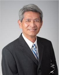
Julin F Tang
Zuckerberg San Francisco General Hospital and Trauma Center, USA
Keynote: Unexpected right to left shunt of air embolism during cardiopulmonary resuscitation of a patient undergone endoscopic retrograde cholangiopancreatography
Time : 10:30-11:15

Biography:
Julin F Tang has completed his MD, MS & FCCM and is a HS Clinical Professor in Anesthesia & Perioperative Care University of California, San Francisco, School of Medicine. His areas of interests include cardiopulmonary physiology, echocardiography, transesophageal echocardiography, traumatic brain injury, critical care, respiratory care, fluid resuscitation, conscious sedation, neuromuscular problems, airway management, acidosis, alpha2 agonist, trishydroxyaminomethane (THAM), aerosolized prostacyclin, mechanical ventilation, trauma, geriatric anesthesia, ventilator associated pneumonia, artificial breathing patterns, capnography, bronchoalveolar lavage, methicillin-resistant Staphylococcus aureus, cardiac ultrasonography and lung scan.
Abstract:
Keynote Forum
Agnes Molnar
Westmead Private Hospital, Australia
Keynote: Unrecognized anaphylaxis under anesthesia
Time : 11:30-12:15

Biography:
Agnes Molnar is a Specialist Anesthesiologist in Sydney, Australia. She has completed her Medical studies and gained specialist qualification in Anaesthesia and Intensive Therapy first in her native Hungary. Following her husband, she migrated to Australia and started a long journey to become an Anesthetist in her adopted country. She works as a Visiting Medical Officer in Western Sydney in both the public and the private system. She has extensive experience with Bariatric Patients.
Abstract:
Case of a morbidly obese 45 year old woman (BMI 50) undergoing a minor day case procedure with planned general anaesthesia and intubation who, after a standard induction including suxamethonium, presented a challenging airway and bronchospasm simultaneously. Haemodynamics initially were uncompromised with the main issues being sudden onset significant desaturation and bronchospasm which was managed after intubation with endotracheal salbutamol. Skilled assistance was sought from anaesthetic and intensive care colleagues. Rocuronium administration was then required after successful intubation had been confirmed and after saturations had improved, to facilitate further stabilization when the patient started to wake and cough on the endotracheal tube. After rocuronium was administered the ongoing bronchospasm and desaturation worsened again with subsequent haemodynamic instability. The patient’s body habitus made NIBP monitoring unreliable and difficult intravenous access further hindered attempts to fluid resuscitate, necessitating insertion of an arterial line and central venous catheter. Small boluses of adrenaline were administered for low blood pressure and in view of ongoing instability it was decided that surgery should be abandoned, and she was transferred to the intensive care unit intubated, where she was managed with a noradrenaline infusion. Mast Cell Tryptase was sent for suspected anaphylaxis after 28 hours when attempts at weaning from the ventilator proved difficult, despite having been haemodynamically stable and off vasopressors for more than 12 hours. Mast Cell Tryptase came back elevated and subsequent formal allergy testing confirmed a positive reaction to both suxamethonium and rocuronium. Despite quite a brief period of significant desaturation the patient unfortunately sustained hypoxic brain injury, although she has been able to return home and her cognitive function continues to improve. The case highlights the difficulties in managing emergency scenarios in situations where; More than one crisis occurs simultaneously (difficult airway management and anaphylaxis presenting initially with isolated bronchospasm); Morbidly obese patients present specific challenges regards to monitoring and iv access, delaying resuscitation efforts. Team leadership is not clearly defined in a scenario where several equally qualified experts are present.
Keynote Forum
Mohamed Farouk Helal
Miltary Medical Academy, Egypt
Keynote: Synchronous double primary cancer of the frontal sinus and the nasopharynx: A case report and review of the literature
Time : 12:15-13:00

Biography:
Mohamed Farouk Helal is an Associate Professor of ENT at Military Medical Academy, Egypt. Currently, he is ENT Consultant at Dallah Hospital, KSA. He has a special interest in Endoscopic Sinus Surgery. He was an Ex-Major General Dr. at Egyptian Armed Forces, was Winner of Long Service and received good example medal from Mr. President of Egypt 2007.
Abstract:
We present a case report of a 55 years old male patient who has been diagnosed as having Inverted papilloma of the frontal sinus which was incompletely removed elsewhere in 2014. The patient main complaint was nasal obstruction which improved after removing that part of the IP presenting in the nose. The patient's symptom of nasal obstruction recurred few months after his first surgery but he ignored it till few months ago when he started to have frontal headache and right sided proptosis moreover he complains of bilateral blocked ears which were diagnosed as bilateral otitis media with effusion. Radiologic workup (CT and MRI) showed erosion of the posterior table of the frontal sinus and the roof of the right orbit with infiltration of the Dura by the mass already left over from the previous surgery and diagnosed as IP. The nasopharynx showed a huge mass abutting the fossa of Rosenmullar and the eustachian tube bilaterally with suspicious retropharyngeal lymph nodes. This mass was found to be both radiological and surgically distinct from the frontal sinus mass. Histopathology revealed an inverted papilloma with malignant transformation involving the Dura of the floor of the anterior cranial fossa and a synchronous second nasopharyngeal carcinoma. The patient was referred to oncology department for combined chemo and radiotherapy.
Keynote Forum
Samih Abed Odhaib
Faihaa Specialized Diabetes Endocrine and Metabolism Center, Iraq
Keynote: How biotin induces thyroid function tests misleading results: case series

Biography:
Samih Abed Odhaib has completed his Medical Education from Nahrain Medical School in 2001. He is a Fellow of Iraqi Board of Medical Specialization (Internal Medicine) FIBMS, Member of American College of Physician (ACP), Member of American Association of Clinical Endocrinologist (AACE). He is a Post-doctorate in Adult Endocrinology and is associated with Faiha Specialized Diabetes, Endocrine, and Metabolism Center (FDEMC) since September 2018. He was the Head of the Study Group that manage the case of Right Paraduodenal Hernia and the Manuscript Author.
Abstract:
Introduction: We present four cases that show a misleading increase in the thyroid function test towards a false diagnosis of Grave's disease after biotin administration of 20-30 mg for different periods, although all patients are free of signs and symptoms of hyperthyroidism, and all the results returned to normal after biotin withdrawal within 24-48 hours.
Case 1: A 23-year-old female was diagnosed as Grave's disease after a routine checking after she ingested prescribed 20 mg biotin per day for three months for excessive hair fall.
Case 2: A 19-year-old female with hair and nail problems associated with iron deficiency anemia. She had self-prescribed biotin dose of 20 mg a day for a month. She asked an endocrinologist opinion about a recent increase in her thyroid function tests, yet no signs of hyperthyroidism were found.
Case 3: A 45-year-old man with subtotal thyroidectomy for a retrosternal multinodular goiter with compressive symptoms, his usual levothyroxine dose had been decreased from 100 to 50 microgram per day, after which he felt unwell and gained four kilograms with signs and symptoms of hypothyroidism. His investigations were consistent with hyperthyroidism while his signs were of hypothyroidism which was illogical. He administered 30 mg medically prescribed biotin for a recent mild psoriasis nail changes.
Case 4: The author himself volunteered to take 30 mg of biotin daily for one week. His initial investigations were normal but changed within this period to be Grave's disease-like, with no signs or symptoms to be felt.
Keynote Forum
Mohamed Farouk Helal
Miltary Medical Academy, Egypt
Keynote: Synchronous double primary cancer of the frontal sinus and the nasopharynx: A ca report and review of the literaturese

Biography:
Mohamed Farouk Helal is an Associate Professor of ENT at Military Medical Academy, Egypt. Currently, he is ENT Consultant at Dallah Hospital, KSA. He has a special interest in Endoscopic Sinus Surgery. He was an Ex-Major General Dr. at Egyptian Armed Forces, was Winner of Long Service and received good example medal from Mr. President of Egypt 2007.
Abstract:
We present a case report of a 55 years old male patient who has been diagnosed as having Inverted papilloma of the frontal sinus which was incompletely removed elsewhere in 2014. The patient main complaint was nasal obstruction which improved after removing that part of the IP presenting in the nose. The patient's symptom of nasal obstruction recurred few months after his first surgery but he ignored it till few months ago when he started to have frontal headache and right sided proptosis moreover he complains of bilateral blocked ears which were diagnosed as bilateral otitis media with effusion. Radiologic workup (CT and MRI) showed erosion of the posterior table of the frontal sinus and the roof of the right orbit with infiltration of the Dura by the mass already left over from the previous surgery and diagnosed as IP. The nasopharynx showed a huge mass abutting the fossa of Rosenmullar and the eustachian tube bilaterally with suspicious retropharyngeal lymph nodes. This mass was found to be both radiological and surgically distinct from the frontal sinus mass. Histopathology revealed an inverted papilloma with malignant transformation involving the Dura of the floor of the anterior cranial fossa and a synchronous second nasopharyngeal carcinoma. The patient was referred to oncology department for combined chemo and radiotherapy.
- Colorectal surgery case reports,Cardiology case reports,Gastroentrology case reports,Neurology case reports,Pulmonology case reports,Anaesthesiology case reports,Surgery case reports,Case reports on adverse drug reactions
Session Introduction
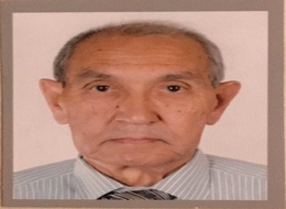
Karimbayev Kidirali
International Kazakh-Turkish University, Kazakhstan
Title: Late and rare complications of urolithiasis: Paranephric abscess and renal replacement lipomatosis

Biography:
Karimbayev Kidirali graduated from Tashkent Medical Institute as a Bachelor of Medicine in 1969, worked in regional hospital in Uzbekistan as an Anesthesiologist during 1969-1976 and in the year 1977 specialized as an Urologist. He earned Post-graduate degree in Urology at the Central Post Graduation Institute (Moscow 1977-1980) and worked as an Assistant Professor at the Department of Urology of Tashkent Medical Institute (1980-1993), then Head of Department of Urology of Republic Hospital (Tashkent, Uzbekistan). Since September 2013, he is working as Professor of Urology in Medical School of International Kazakh-Turkish University. In 1981, he completed his Dissertation Research Work “Transurethral resection in diagnostics of prostatic cancer” and was awarded with the degree of PhD and in 2002 - Dissertation Research Work “Modern methods in the treatment of long and complicated urethral strictures” Moscow with degree of Doctor of Medicine. He has more than 60 articles which were published in republican and international journals. Also, he is a Translator of the “Robbins Basic Pathology”. He translated the book from English to Kazakh language to give Kazakh Medical Students opportunity to study foreign Medical literature.
Abstract:
Background: We present a case of 62 year-old Kazakh women who for a long period of time suffered from back pain, weakness (has been treated with osteochondrosis), and was not aware about her kidney stones. She had never had hematuria or renal colic and urgently was admitted in a severe condition with large palpable abdominal mass.
Methods: Physical examination, laboratory data, radiology of the urinary tract, surgical treatment, histology, medical follow-up.
Results: On physical examination: The lumbar region on the right side is oedematic, hyperemic, and sharply painful on palpation. Oedema spreads down into iliac region and suprapubic area; and medially – to the median line with involvement of the umbilical ring. MRI of the urinary tract: The size of the right kidney is reduced; shadow of multiple stones seen in; the parenchyma is thinned. Beginning from the lower pole of the right kidney up to the pelvic cavity – shadow of a liquid mass with heterogenous density, measuring 21 x 8.4 cm is determined.
Surgical Procedure: Lumbotomy, lower paranephral abscess drainage. Later – right nephrectomy.
Histology: Renal replacement lipomatosis, severe renal tubular dystrophy.
Conclusions: Nontreated urolithiasis may lead to loss of the kidney function and development of life-threatening complications like paranephric abscess and renal replacement lipomatosis.

Biography:
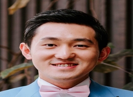
Tony Ze Li is a General Surgical Registrar at The Canberra Hospital with a keen interest in Vascular Surgery and Cardiothoracic Surgery. He was a graduate from the prestigious Australian National University and has stayed locally to work in the Australian capital region since then. He has also done cardiac surgical placements in Texas Heart Institute in Houston, Texas. He is particularly interested in the endovascular aspects of vascular surgery based on the combination of both the surgical finesse of the procedures as well as the ever changing technology involved in this field. Before his career in Medicine, he has attained university degrees in both Biomedical Science and Biotechnology Commercialization.
Abstract:
Background: Endovascular aneurysm repair (EVAR) has been a long-stay surgical option to treat abdominal aortic aneurysms (AAA), both contained and ruptured. Endoleaks, defined as persistent flow in the aneurysm sac extrinsic to the endograft, is the most common complication. Type II endoleak (T2EL) results from collateral aortic branches (lumbar arteries, inferior mesenteric artery) flowing retrograde into the sac. It is often considered the Achilles' heel to EVAR due to the controversies in its timing of diagnosis and management. It’s accepted that T2EL are slow growing, could expand the aneurysm sac, and carry a small but significant risk of aneurysm rupture.
Case Presentation: This case reports an early post-EVAR complication due to T2EL. Sixty six (66) years old gentleman was brought to casualty hypotensive, minimally responsive with a rigid and tender abdomen. CT angiography revealed a ruptured AAA contained within the retroperitoneum. The patient subsequently underwent an uncomplicated EVAR procedure. However one hour post procedure in ICU, he was found to be suddenly in haemorrhagic shock. Exploratory laparotomy revealed 8 L of blood within the peritoneum. Aneurysm sac was opened and four strong bleeding lumbar arteries were found and oversewn. It’s hypothesized that vigorous T2EL from lumbar back- bleed, through the ruptured aneurysm sac led to ongoing bleeding into the retro-peritoneum and subsequently into the peritoneal space.
Conclusion: This case showed that T2EL are not all slow growing and innocuous. One should consider T2EL as a cause of a patient who’s acutely deteriorating post-EVAR. Early CT-angiographic imaging post procedure may be indicated in certain groups.
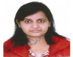
Shweta Dixit
Sir Gangaram Hospital, India
Title: The Etiology outcome and prognostic indicators in children with acute hepatic failure

Biography:
Shweta Dixit is presently pursuing a fellowship in Pediatric Gastroenterology from Sir Gangaram Hospital, New Delhi. She has done Diploma in Child Health and DNB in Pediatrics.
Abstract:
Objective: To study the etiology, outcome and prognostic indicators in pediatric acute liver failure (ALF).
Material & Method: A retrospective analysis of patients with ALF between 2010 and 2018 was performed. There were total 90 patients. Patients who survived with supportive therapy were designated as Group 1, while those who died or required liver transplantation were designated as Group 2.
Results: There were 90 children- 64 male, 26 females, median age eight years; range, one month-17 years were identified with fulminant hepatic failure. In Group 1 there were 53 (58.9%) patients and 37 (41.1%) patients in Group 2. The overall survival rate was 74%. Eleven (11) patients (12.2%) underwent liver transplantation, all are alive and well. The etiologies were: 60 (67%) infectious, 5 (5.5%) drug-induced, 5 (5.5%) Wilson disease and 3 (3.3%) autoimmune hepatitis. Overall acute viral hepatitis A is the most common cause. There were 16 (17.8%) cases which were of indeterminate etiology. Group 2 children had higher plasma bilirubin (median, 18.4 mg/dl versus 8.8 mg/dl), higher prothrombin time (median, 50.3 seconds versus 31.7 seconds) higher international normalized ratio (INR) (median, 4.9 vs 2.9) lower alanine aminotransferase (median, 1128 IU/L versus 2211 IU/L) and lower aspartate aminotransferase (median, 640 IU/L versus 1839 IU/L) levels on admission (P<0.005). Using receiver operating characteristic curves the cutoff values as predictor of the eventual failure of conservative therapy were serum bilirubin >13.6 mg/dl, prothrombin time >35.8 sec, INR >3.7, alanine aminotransferase ≤1913 IU/L, aspartate aminotransferase ≤ 1744 IU/L on admission.
Conclusions: Pediatric acute liver failure patients with high bilirubin, severe coagulopathy, lower alanine aminotransferase and lower aspartate aminotransferase level on admission are more likely to have poor prognosis. Early consideration of liver transplantation is of paramount importance in these patients.
Samih Abed Odhaib
Faiha Specialized Diabetes, Endocrine, and Metabolism Center,Iraq
Title: Preoperative diagnosis of a rare small bowel obstruction : Right paraduodenal herina: Case Report

Biography:
Samih Abed Odhaib has completed his Medical Education from Nahrain Medical School in 2001. He is a Fellow of Iraqi Board of Medical Specialization (Internal Medicine) FIBMS, Member of American College of Physician (ACP), Member of American Association of Clinical Endocrinologist (AACE). He is a Post-doctorate in Adult Endocrinology and is associated with Faiha Specialized Diabetes, Endocrine, and Metabolism Center (FDEMC) since September 2018. He was the Head of the Study Group that manage the case of Right Paraduodenal Hernia and the Manuscript Author.
Abstract:
The right paraduodenal hernia is an internal hernia of a rare type extends through the fossa of Waldeyer, caused by a congenital gut malrotation defect or trauma, infectious disease, iatrogenic trauma or mesenteric defect. The right is far less common than the left paraduodenal hernia (1:3), and three times more male predominant. A 67-year-old female with acute small bowel obstruction, with a diagnosis of right paraduodenal hernia done, preoperatively by imaging studies and confirmed during laparotomy with a good postoperative course and total resolution of obstructive symptoms. This will be the 168th case reported since Neubauer had published his first case in 1756.
